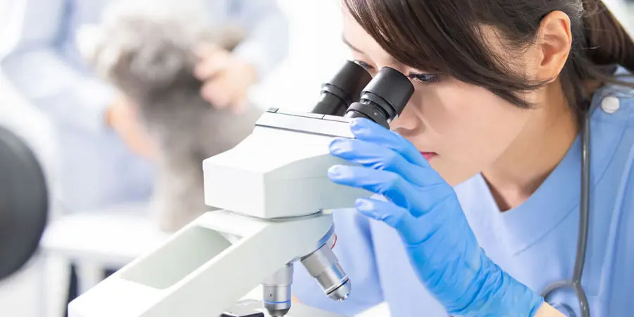
The Role of Microscopes in Modern Medicine and Research
The human body holds a universe of tiny wonders. Cells, pathogens, and fine structures remain hidden from our bare eyes. For many years, our understanding of health and sickness was limited by what we could see. Then came the microscope. This amazing tool broke those limits. It opened a window into the world of cells and molecules. This key technology has changed how we diagnose problems, find treatments, and understand life itself.
From finding the germs that cause sickness to mapping our genes, microscopes do so much. They even guide careful surgeries. The microscope’s effect on medicine and research runs deep. It is more than just a tool. It pushes progress forward. It keeps showing us smaller details, opening new paths for science and patient care.
Unveiling the Microscopic World: A Brief History of Microscopy in Medicine
Early Innovations and the Dawn of Cellular Biology
The journey of the microscope started centuries ago. Zacharias Janssen likely made the first compound microscope around 1590. This early device brought tiny things into view. Later, Antonie van Leeuwenhoek made his own simple microscopes. He was the first to see and draw bacteria, protists, and blood cells. His work showed us that a world of tiny living things existed all around us. This early work laid the groundwork for understanding what living things are truly made of.
Advancements in Light Microscopy: Resolution and Contrast
Scientists quickly worked to make microscopes better. They improved lens designs. New ways to light up samples, like Köhler illumination, helped immensely. Robert Hooke famously used a microscope to see "cells" in cork in 1665. Stains also played a big role. Gram staining, for example, helped tell different bacteria apart. These steps greatly improved how clearly we could see cell parts and tiny germs.
Beyond Light: Electron Microscopy and its Transformative Power
Light microscopes had a limit to what they could see. In the 1930s, the electron microscope appeared. It used beams of electrons instead of light. This new tool offered much higher power. Transmission Electron Microscopes (TEM) let us see tiny parts inside cells. Scanning Electron Microscopes (SEM) showed detailed 3D surfaces. Electron microscopes transformed how we understood viruses and cell structures.
Microscopes in Diagnostics: Pinpointing Disease with Precision
Identifying Pathogens: From Bacteria to Viruses
Microscopes are vital for finding what makes people sick. Doctors use them daily to spot bacteria, fungi, and parasites. They look at samples like blood, tissue, or urine. For example, a doctor might use a microscope to find the bacteria that causes tuberculosis. Or they might look for malaria parasites in a blood smear. Seeing these tiny invaders helps doctors choose the right medicine fast.
Cellular Pathology: Cancer Detection and Blood Analysis
Microscopy is key in diagnosing serious diseases like cancer. Pathologists examine tissue samples under a microscope. They look for abnormal cells. This helps them diagnose cancer, decide its type, and see how far it has spread. In blood tests, microscopes help check blood cell shapes and numbers. They can spot signs of anemia or leukemia. Papanicolaou (Pap) stains on cell samples are also vital for cervical cancer screening.
Emerging Diagnostic Applications: Real-time and Advanced Imaging
Newer microscope methods are speeding up diagnoses. Digital microscopes allow images to be shared and analyzed easily. Live-cell imaging lets doctors watch changes in cells as they happen. These tools help with quick diagnoses. They also help track if a treatment is working over time. This means faster and more accurate care for patients.
Microscopes in Therapeutic Development: Guiding Drug Discovery and Treatment
Screening and Identifying Drug Targets
Microscopes play a big part in finding new medicines. Scientists use them in high-throughput screening. This lets them test many drug compounds at once. They watch to see how these compounds affect cells or disease models. By looking through the microscope, they can see if a drug changes cells in the right way. This helps find promising drug candidates.
Understanding Drug Mechanisms of Action
Advanced microscopes help us learn how drugs work. Techniques like fluorescence and confocal microscopy let scientists track drugs. They can see where a drug goes inside a cell. They can also see which cell pathways a drug affects. This helps researchers understand why a drug works. It also helps them figure out why some drugs stop working, which we call drug resistance.
Minimally Invasive Surgery and Targeted Therapies
Microscopes are not just for labs. They are critical tools in modern surgery. Surgeons use special microscopes during delicate operations. This is common in neurosurgery or eye surgery. These surgical microscopes provide a magnified, clear view. This allows surgeons to work with extreme precision. They can protect healthy tissue and target only the problem area.
Cutting-Edge Microscopy Techniques Shaping the Future of Research
Super-Resolution Microscopy: Breaking the Diffraction Limit
Regular light microscopes have a limit to how small they can see. Super-resolution microscopy breaks this barrier. Techniques like STED, STORM, and PALM allow us to see things previously too small. They show molecular interactions in amazing detail. This helps scientists understand how proteins work inside cells. It opens up new ways to study life at its most basic level.
Advanced Live-Cell Imaging and Functional Assays
Watching living cells in real-time gives us key insights. Live-cell imaging lets scientists study how cells move, talk to each other, and how their tiny parts work. Techniques like Förster Resonance Energy Transfer (FRET) show when molecules interact directly. This helps us see how dynamic our cells truly are. It reveals the complex dance of life.
Integration with AI and Machine Learning
The future of microscopy is getting smarter. Artificial intelligence (AI) and machine learning are now used with microscopes. These tools can automatically analyze images. They find complex patterns in large sets of data. This speeds up drug discovery. It also helps classify diseases more quickly and accurately. AI helps us get more from every image.
Conclusion: A Continued Legacy of Vision and Discovery
The microscope started as a simple idea. Today, it is a complex, vital tool. It has always pushed medicine and research forward. It has given scientists and doctors the power to see, understand, and fight illnesses that once seemed like mysteries. As technology keeps improving, especially with AI and new ways of seeing, the microscope's role will only grow. It promises even greater strides in diagnoses, treatments, and our basic understanding of life.
Seeing the microscopic world remains hugely important. It offers endless chances for new ideas and better results for patients.


